
Skeletal Muscles
Skeletal muscles connect to bones either by tendons or aponeurosis and movement occurs when they contract. The origin of a muscle is the bone to which it attaches, the origin does not move when it contracts whereas the point of attachment is the bone that moves when the muscle contracts.
In order to facilitate movement, skeletal muscles come in antagonistic pairs; this means that when one contracts, the antagonist relaxes to allow for opposing movements.
Skeletal muscles enable the movement of the body in space by acting on joints. Some examples of these movements include:

Flexion and Extension - bending and straightening
Abduction and Adduction - moving away from and towards the midline of the body
Pronation and Supination - rotating the forearm such that the palm faces the ground or the ceiling
Elevation and Depression - moving a particular body part upwards or downwards
Protraction and Retraction - moving a bone forward or backward along the same plane
Inversion and Eversion - turning the soles inward or outward
Dorsiflexion and Plantarflexion - pointing the toes towards the shins or the floor.
Types of Skeletal Muscles
Circular - e.g. orbicularis oris
Convergent - e.g. pectoralis major
Multipennate - e.g. deltoid
Parallel - e.g. biceps brachii
Unipennate - e.g. extensor digitorum
Bipennate - e.g. rectus femoris
Structure of a Skeletal Muscle
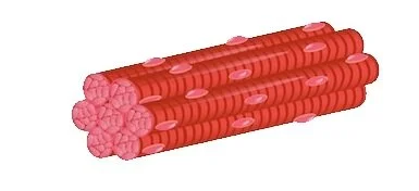
Every skeletal muscle is bundled together by three layers of connective tissue, the epimysium, perimysium and endomysium.
Epimysium: This is the outermost later of irregular connective tissue that allows the muscle to contract and move independently from other structures.
Perimysium: This is the middle layer of connective tissue that holds together bundles of muscle fibers called fascicles. These fascicles allow movements to take place by activating the muscle fibers within them when triggered by the nervous system.
Endomysium: This innermost layer of connective tissue can be found around each muscle fiber found in the fascicles.
All skeletal muscles are generously supplied by blood vessels and by the axon branch of a somatic motor neuron which signals the fibers to contract.
Myofibrils
Myofibrils have many nuclei, mitochondria and the following specialized organelles:
Sarcolemma - the plasma membrane of muscle fibers
Sarcoplam - the cytoplasm of muscle fibers
Sarcoplasmic reticulum - there organelles store, release and retrieve calcium ions
Sarcomere - smallest functional unit of a skeletal muscle fiber, contains contractile, regulatory and structural proteins
These myofibrils are made up of proteins and span the length of the cell and they contain thousands of sarcomeres.
When each individual sarcomere contracts, it results in the contraction of the muscle fiber that it resides in. The contraction of all the individual sarcomeres leads to the entire muscle contracting.
What is a sarcomere and how does it function?
Sarcomere
Sarcomeres are made up of two protein myofilaments:
Thick filament - these are called myosin and they contains small heads that bind to the thin filament
Thin filament - these are called actin to which the myosin binds
The continuous binding of myosin heads to actin filaments and its subsequent release, lead to the constant lengthening and shortening of each sarcomere.
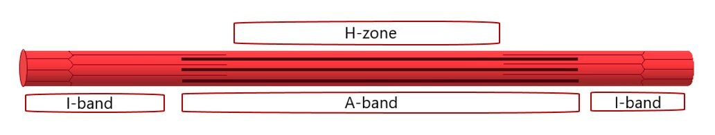
Sarcomeres are contained in the region within two Z-lines. Actin filaments fan out form the Z-lines and help to hold myofilaments down in place. The recurring pattern of sarcomeres along each myofibril create its striated appearance.
A-band: Where actin and myosin filaments overlap each other
I-band: Region where only actin filaments are present in the sarcomere
H-zone: regions of the A-band where only myosin filaments are present
How do skeletal muscles contract?
Muscle contraction can be summarized into four key steps:
Depolarisation
When a motor neuron receives an action potential, it triggers the release of acetylycholine. In the sarcolemma, this initiates the process of depolarisation. Depolarisation then causes the sarcoplasmic reticulum to release the reserves of calcium ions within it. These calcium ions then intiate muscle contractions.
2. Bridge Formation
Troponin and tropomysin are little proteins that are locked on actin filaments which block myosin heads from binding to them. When calcium ions are released, they begin to bind with troponin which opens up their position on the actin filaments, allowing myosin heads to bind to it. This binding of myosin heads and actin filaments forms a bridge.
3. Sliding Mechanism
The bridges that were previously formed, now get broken up by adenosine triphosphate. When adenosine triphosphate hydrolyses, it helps to move the myosin heads towards another actin filament where it once again binds to. This movement creates a sliding mechanism.
4. Shortening of Sarcomere
Actin filaments are firmly bound to the Z-lines and each time a myosin head moves to bind to a new site, the Z-lines are pulled together. This in turn shortens the individual sarcomere which in turn contracts the muscle fiber.

Please refer to the ‘Anatomy By Regions’ tab for specific skeletal muscles in each region